
Description
Aim
…at enhancing patient outcomes
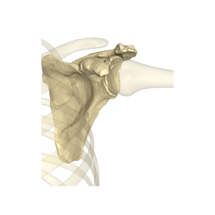
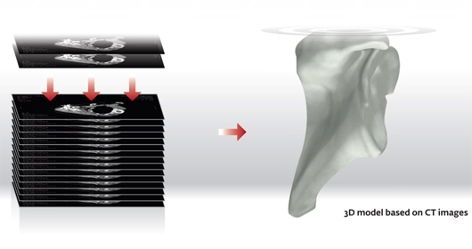
Starting with a preoperative CT scan, surgeons can Aim for improved patient outcomes. The CT scan is uploaded into proprietary software in which a 3D model of the patient’s shoulder is rendered. This allows surgeons to visualize the patient’s unique anatomy on a computer, focusing on the patient’s specific condition in ways not possible during conventional surgery. Having the ability to preoperatively view the patient’s anatomy can help prepare the surgeon to handle any nuances that could otherwise be a surprise in surgery.
Set
…patient goals to Reach Higher
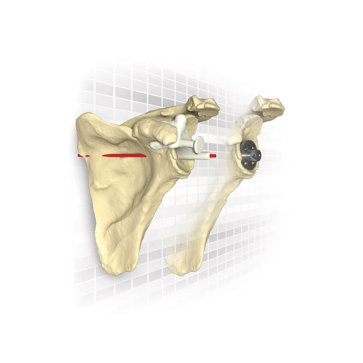
By virtually aligning the implant specifically to the patient’s anatomy, surgeons can preoperatively Set the optimal position for the implant, taking full advantage of the design and potentially improving the implant performance. Surgeons can feel confident going into surgery with a plan optimized for the patient which will help reduce the complications that can arise from unforeseen bone morphology.
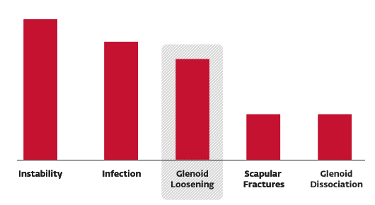
Aseptic loosening of the glenoid component is one of the leading causes of postoperative complications in both total and reverse shoulder arthroplasty.1,2
Failing to restore the glenoid version angle has been cited to place the implant at increased risk of premature loosening.3 Studies have shown traditional surgery methods are not reproducible at restoring the retroversion angle of a severely deformed glenoid (B2).3,4
Studies have shown the correct placement of the glenoid component is essential for:
Postoperative Complications1
- Longevity of the prosthesis3
- Positive effects on functional outcomes3
Matched
…to the patient’s specific anatomy

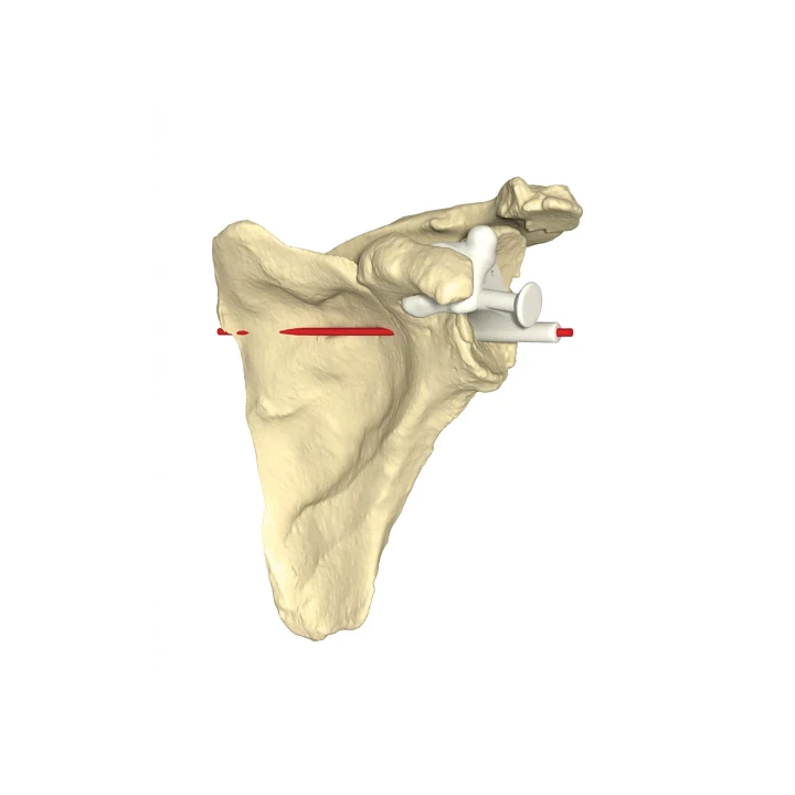
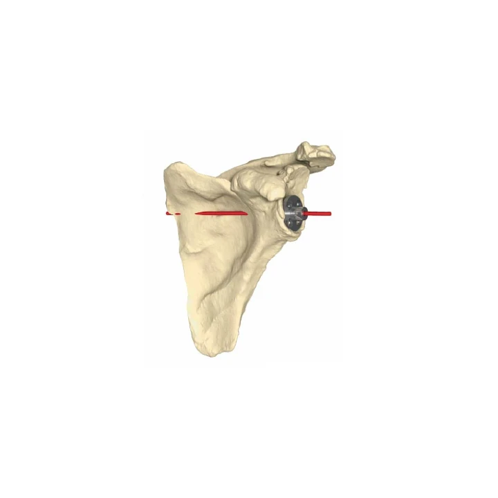
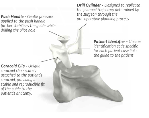
Conventional implantation methods have been shown to be highly variable, with implant inferior tilt and version angles in excess of 16° and 12°, respectively, of the planned alignment.5,6 With the Match Point System® from DJO Surgical®, the unique coracoid clip attaches securely to the patient’s anatomy in surgery, allowing for a more accurate placement of the surgical implant.
The Match Point System guides provide a drill path validated to replicate the preoperatively planned position on average within 2.5° for glenoid version and 1.5° for inferior tilt.7
The Set plan is used to design and manufacture a guide and model to be used by the surgeon during the procedure. The guide is Matched to the patient’s anatomy to ensure secure attachment to the bone. Once in position, the surgeon drills through the guide, so what was planned on the computer is Matched to the patient’s actual bone.
Reviews
There are no reviews yet.